From today’s West Australian.
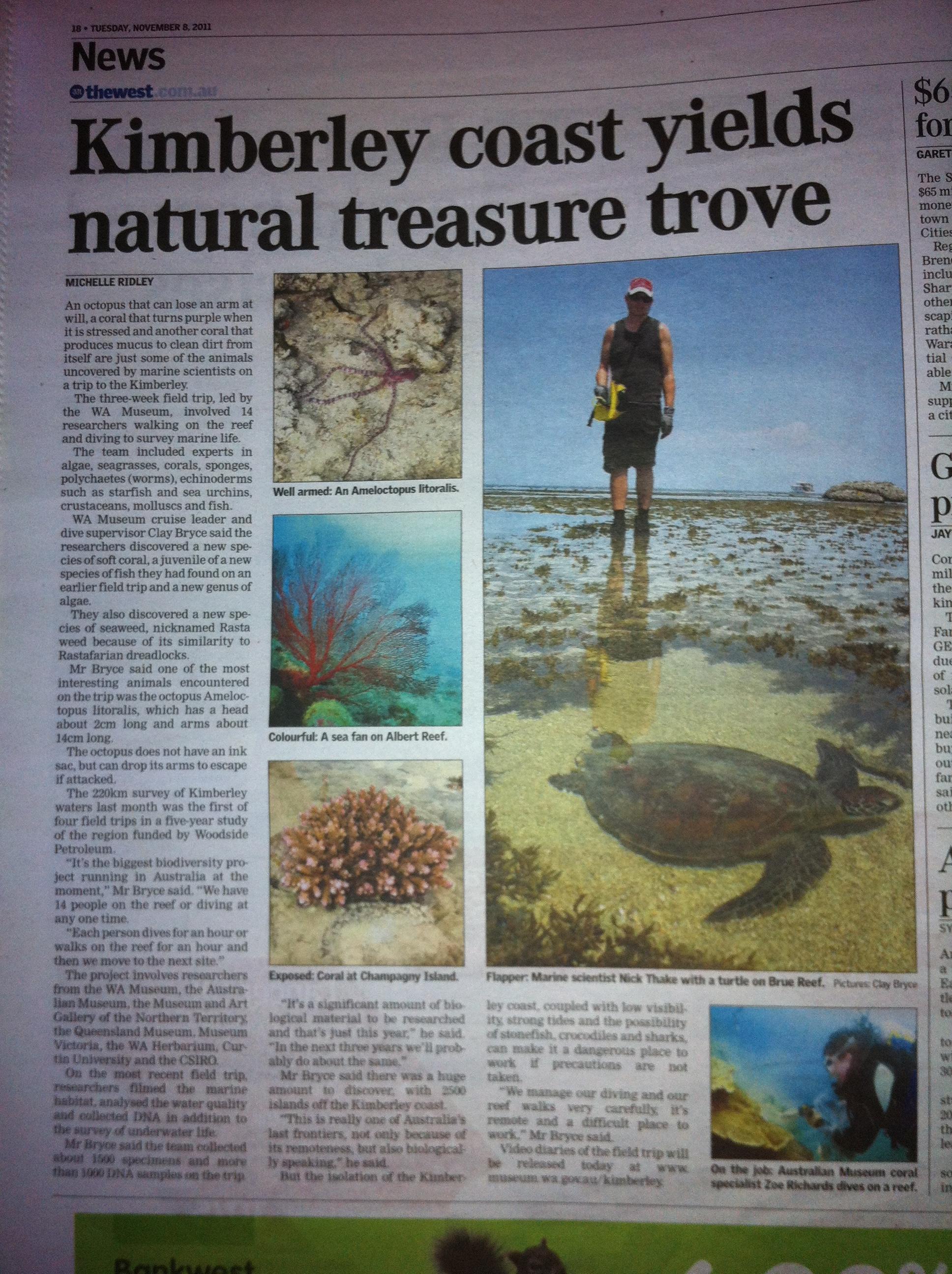
Check out new aqua critters in the Kimberley coast
From today’s West Australian.
All about Fish Vetting – Dr Richmond Loh
From today’s West Australian.
Begin forwarded message:
From: noreply>
Date: 8 November 2011 6:20:06 AWST
To: Undisclosed Recipients
Subject: Disease News Update
AquaticHealth.net Disease News Update >> Catfish tumor – USA [ProMed request for information]
Link to source content: http://www.hometownannapolis.com/news/TOP/2011/10/31-31/Cause-of-catfish-tumors-still-unknown.html.
Your input is important: Please verify and add any relevant commentary to this report. Also, is the information in the report summarised somewhere in the Wiki?
Report Content:
CATFISH TUMOR – USA: (MARYLAND) REQUEST FOR INFORMATION
*******************************************************
A ProMED-mail post
ProMED-mail is a program of the
International Society for Infectious Diseases
Date: Mon 31 Oct 2011
Source: Howntownannapolis.com, The Capital Gazette [edited]
Cause of catfish tumors still unknown
Aquaculture International: Journal of the European Aquaculture Society Volume 19, Number 6 (December 2011) 
Ovaprim treatment promotes oocyte development and milt fertilization rate in diploid and triploid African catfish (Clarias gariepinus)
Authors: Ali Karami 1, Annie Christianus 1, Hadi Zokaeifar 1, Khairul Saad 1, Fahmi Imraan 1, Shahram Shakibazadeh 1, Hossien Negarestan 2, Simon Courtenay 3 Author Affiliations:
| 1: | Department of Aquaculture, Faculty of Agriculture, University Putra Malaysia, 43400 Selangor, Malaysia |
| 2: | Department of Aquatic Ecology of Caspian Sea, Iranian Fisheries Research Organization (IFRO), Tehran, Iran |
| 3: | Fisheries and Oceans Canada at the Canadian Rivers Institute, Department of Biology, University of New Brunswick, Fredericton, NB, Canada |
Source: Aquaculture International: Journal of the European Aquaculture Society, Volume 19, Number 6 (December 2011) Page Numbers: 1025 – 1034 Available Full Text:
| Full Text: | Subscription Required to view full text |
| Format: | |
| Size: | unknown |
| Location: | Publisher’s Site |
| Authentication: | EBSCOhost EJS |
Abstract:
Citation: Ali Karami, Annie Christianus, Hadi Zokaeifar, Khairul Saad, Fahmi Imraan, Shahram Shakibazadeh, Hossien Negarestan, Simon Courtenay . Ovaprim treatment promotes oocyte development and milt fertilization rate in diploid and triploid African catfish (Clarias gariepinus). Aquaculture International: Journal of the European Aquaculture Society, Volume 19, Number 6 (December 2011), pp. 1025-1034,
Keeping it simple….
From today’s West Australian.

From today’s West Australian.
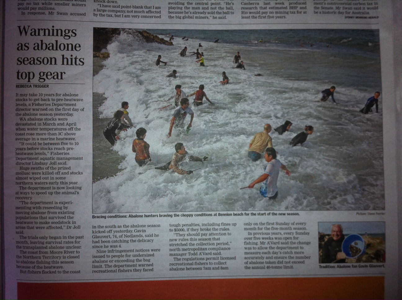
Aquaculture Volume 321, Number 3-4 (December 2011)
Organic acids as potential growth promoters in the South African abalone Haliotis midae
Authors: Neill Jurgens Goosen, Johann Ferdinand Görgens, Lourens Francois De Wet, Hafizah Chenia Author Affiliations:
| no affiliations available |
Source: Aquaculture, Volume 321, Number 3-4 (December 2011) Page Numbers: 245 – 251 Available Full Text:
| Full Text: | Subscription Required to view full text |
| Format: | |
| Size: | Unknown |
| Location: | Publisher’s Site |
| Authentication: | Publisher’s Site |
Abstract: The study investigated organic acids and organic acid salts as growth promoters in cultured South African abalone Haliotis midaewhen incorporated as feed additives in formulated feed. Two blends of organic acids (1% acetic acid+1% formic acid, and 1% benzoic acid+1% sorbic acid) and one of organic acid salts (1% sodium benzoate+1% potassium sorbate) were evaluated in South African abalone (23mm initial mean length), and tested against a negative control and positive control containing 30ppm avilamycin as antibiotic growth promoter (AGP). Experiments were performed at controlled water temperatures under optimal (Phase 1) and sub-optimal (Phase 2) temperatures. During Phase 1 the organic acid and organic acid salt blends significantly enhanced mass growth rate (both linear weight increase [AGRW] and specific growth rate [SGR]) compared to both controls. Linear length increase (AGRL) was significantly higher than the negative control. SGR was increased by 15.8%–17.9% relative to the negative control; AGRW and AGRL were between 12.9%–22.9% and 11.7%–13.5% higher, respectively. No significant growth enhancement was observed during Phase 2. Avilamycin had no effect on growth and none of the treatments had significant effects on feed conversion ratio (FCR), relative incidence cost (RIC, relative to negative control) or feed intake during either phase. The mode of growth enhancement could not be established; it could not be shown that the acid and salt treatments or the AGP had any effects on intestinal microflora, but possible energy effects of the acids were eliminated as potential mechanism; it is unclear what role gut acidification played in the enhanced growth performance.
Citation: Neill Jurgens Goosen, Johann Ferdinand Görgens, Lourens Francois De Wet, Hafizah Chenia . Organic acids as potential growth promoters in the South African abalone Haliotis midae. Aquaculture, Volume 321, Numbers 3-4 (December 2011), pp. 245-251,
Aquaculture
Volume 321, Number 3-4 (December 2011)
In vitro and in vivo efficacies of ionophores against Cryptocaryon irritans
Authors: T. Yoshinaga, H.J. Im, S. Nishida, K. Ogawa
Author Affiliations: no affiliations available
Source: Aquaculture, Volume 321, Number 3-4 (December 2011)
Page Numbers: 167 – 172
Available Full Text: Full Text: Subscription Required to view full text
Format: PDF
Size: Unknown
Location: Publisher’s Site
Authentication: Publisher’s Site
Abstract: We assayed the effects of a variety of antiprotozoal compounds against trophonts of Cryptocaryon irritans, the causative agent of ‘white spot disease of marine fish’ in vitro using the double layered media that we developed previously for the culture of the parasite. In the assay, ionophores, particularly sodium salinomycin, showed apparent killing and growth-suppression effects against the parasite. As there was no mortality in Japanese flounder Paralychthys olivaceusthat were fed a diet containing sodium salinomycin (200ppm) for two weeks, we evaluated the efficacy of 200ppm sodium salinomycin against C. irritansin Japanese flounder. We fed Japanese flounders a medicated diet for 5d prior to and 3d after challenge with C. irritans.In the experimental group, the number of protomonts recovered from the fish and the size of tomonts that were transformed from the protomonts were significantly reduced, when compared to the control group. Furthermore, in a different experiment, the fish that were fed a diet medicated with sodium salinomycin survived longer than those fed an unmedicated diet after challenge. Sodium salinomycin can be a good candidate drug for chemotherapy and control of Cryptocaron irritansinfection.
Citation: T. Yoshinaga, H.J. Im, S. Nishida, K. Ogawa . In vitro and in vivo efficacies of ionophores against Cryptocaryon irritans. Aquaculture, Volume 321, Numbers 3-4 (December 2011), pp. 167-172,
I wonder if there are parallels with fish tuberculosis…
The article below is from –
http://www.sciencedaily.com/releases/2011/10/111003180430.htm
Tuberculosis bacterium’s outer cell wall disarms the body’s defense to remain infectious
 |
|
|
18 October 2011
|
They first established that human macrophages respond differently to the two different types of bacteria lipomannans after 24 hours of exposure. Lipomannan from the virulent TB bacterium produced significantly less TNF than lipomannan from the M. smegmatis bacterium. Though the study showed that the harmless cells increase production of TNF through a well-known receptor pathway as expected, the virulent TB bacteria did not make use of that receptor pathway. This supported the concept that the pathogenic TB bacterium has figured out another way to block the TNF protein in its quest to keep the immune system guessing, said Schlesinger, also the director of Ohio State’s Center for Microbial Interface Biology. A single microRNA can affect the production of hundreds of proteins, and the process of identifying those relationships is ongoing. However, two microRNAs in this study were known to be relevant for their connections to genes and proteins already established as players in the immune response to TB infection: miR-125b and miR-155.
Biochemical and genetic experiments showed that macrophages stimulated with lipomannan from TB bacteria had enhanced expression of miR-125b, effectively inhibiting the production of TNF. In contrast, the lipomannan from the harmless bacteria had enhanced expression of miR-155, which regulates other compounds in a way that stimulates TNF production. Researchers’ experimental manipulation to lower the expression of miR-125b in macrophages increased production of TNF in response to the TB bacteria lipomannan, further confirming that this regulation of TNF occurred at the microRNA level, Schlesinger said. “This really speaks to the power of the tuberculosis bacterium to adapt to the human host,” he said. “It has had centuries to develop a sophisticated way to deal with its encounter with the human. Fortunately, genomic technology is allowing us to identify microRNAs more and more rapidly, which might allow us to catch up with the TB bacterium and figure out a way to outsmart it.”
