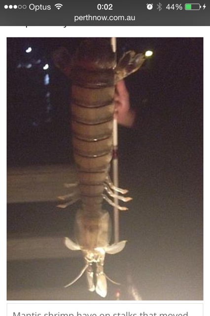
Massive mantis shrimp caught by Florida fisherman stumps scientists.

http://m.perthnow.com.au/technology/science/massive-shrimp-caught-by-florida-fisherman-stumps-scientists/story-fnjwk7lb-1227050266817?nk=0475b03cac14dea94393fbf632777250

—
Yours sincerely,

Dr Richmond Loh
DipProjMgt, BSc, BVMS, MPhil (Pathology) Murdoch, MANZCVS (Aquatics& Pathobiology), CertAqV, CMAVA, NATA Signatory.
THE FISH VET, Perth, Western Australia.
Veterinary Medicine for fish.
W: http://www.thefishvet.com.au
E: thefishvet
P: +61 (0)421 822 383

How can the veterinary pathologist beat the fish bacteriologist to a diagnosis?

BACTERIAL PATHOGENS IN FISH

At last week’s AAPSP Workshop on Fish Pathology, Dr Judith Handlinger provided a beautiful summary of the intricacies in identifying the bacterial disease agents based on observable microscopic lesions. I loved this section of the workshop the most. Imagine that you can beat the bacteriologist to the diagnosis! But in reality, pathologists rely on multiple lines of evidence for a definitive diagnosis.


Gram negative bacteria

Aeromonas salmonicida tend to occur as multifocal dense colonies of fine Gram negative bacteria, free within blood vessels, and in the interstitium of kidney in histology. The reason for them to be so dense is because they are non-motile and quite sticky. The atypical strains also show similar pathology, but clinically less severe with lower morbidity and mortality.


In Yersinia ruckeri infection, splenic lesions may include hyperplasia of the macrophage cuffs, fibrin deposits and oedema, and some intralesional, singular, Gram negative, bacterial rods. There is also mild, multifocal, haematopoietic necrosis in the kidney.


Vibrio anguillarum causes severe septicaemia. The spleen can be enlarged and haemorrhage, ellipsoids degenerate (loss of confluent rim of macrophages) and become depleted of lymphocytes (little white pulp remains).


Vibrio harveyi causes enteritis in barramundi and extensive peritonitis. There is a special strain (Vibrio Phenon 53) where you can get the large form of the bacteria on primary cultures, and reversion to small form on subculture. This large and small form has been described in Vibrio cholera. Suspect it might be due to changes in salinity. The large form can be seen encapsulated in bunches, within host cells.


Tenacibaculum maritimum (formerly Flexibacter maritimus) starts as superficial skin erosion. Microscopically, they line up side-by-side and produce expanding waves of necrosis that precede the bacteria. Early stages of bacterial infection, you may see lymphocyte infiltration in the skin. These fish tend to fend of disease. Those that don’t, develop spongy epithelial hyperplasia, epithelial loss, necrosis. In chronic cases, floccular proteinaceous material collect in scale pockets and there is scale necrosis (see osteoclastic resorption). Where it occurs in the gills and fins, there will be exposed cartilage rays.


Flavobacterium columnare (formerly Flexibacter columnaris) infection causes deep skin necrosis, extending into the musculature. Can also get under-running of damage, with bacteria present under the epithelium. Where it occurs in the gills and fins, there will be exposed cartilage rays.


Flavobacterium branchiophilum causes bacterial gill disease, and the lesion they incite is epithelial hyperplasia instead of necrosis. The less toxic the bacteria, the more hyperplasia and secondary lamellar fusion there is. You can see fine bacterial rods on the surface. There is also a lot of mucus production. Beware not to confuse thee bacteria with mucus strands. Perhaps use a Gram stain or Giemsa stain.


Piscirickettsia causes concentric waves of infection/necrosis in the liver and so grossly, you see targetoid patterns. But typically, you see non-specific petechiation of serosa. Microscopically, you may see intracytoplasmic, Gram negative, coccobacilli in histiocytes.


Infections with Photobacterium damselae subsp. Piscicida, you can see aggregates of gram-negative coccobacilli surrounded by Splendore-Hoeppli material.


Epitheliocystis (Chlamydia) occur as dense, intracytoplasmic colonies, in the gill epithelium. It does not seem to cause pathology in the Barramundi, however, there is focal host reaction in the salmon.


Gram positive bacteria

Lactococcus garvieae (previously Enterococcus seriolicida) are Gram positive cocci. They commonly cause peri-orbital congestion, haemorrhage and oedema (hence exophthalmia/pop-eye). Hagemann strain occurs in trout. Cause enlarged ellipsoids in the spleen, reflect increased phagocytic response. On Gram stains, the bacteria are demonstrated within macrophages in splenic ellipsoid cuffs.


Streptococcus iniae you see clumps of bacteria in heart, surrounded by mixed inflammatory cells. The Gram positive cocci are also found in the brain, gills and other blood vessels.


Renibacterium salmoninarum causes bacterial gill disease. It is a Gram positive, diplobacillus, rod shaped, non-motile, non-spore forming, intracellular bacterium. It causes granulomatous interstitial nephritis with myriad intralesional bacteria.


Carnobacterium (Gram-stain-positive rods) and Vagococcus (Gram-stain-positive cocci) infection you get peritonitis. Common in salmonids, post-spawning, due to retrograde infection up the oviduct.


Acid-fast bacteria

Granulomatous responses seen in mycobacteriosis, however, in seahorses it tends to be less granulomatous, and more histiocytic.


Nocardia (Gram positive, acid-fast, filamentous organisms) also evoke a granulomatous response. The bacterial colonies can also be surrounded by Splendore-Hoeppli material.
Technology lending from octopi.

Have you ever though how the octopus is so adept at camouflage?

“Cephalopod skins use a three-layered system for quick camouflage: A top layer of pigmented chromatophores change color in response to signals from underlying muscles and nerves, a middle layer of light-reflecting cells can be turned on and off within seconds, and a bottom layer of white cells, which provide a bright backdrop to control the contrast of patterns. Octopus skin also contains photosensitive cells that detect light and patterns without relying on feedback from the eyes or brain.”

Now engineers have been able to replicate this.

Read more here.

What’s causing feminisation of fish in rivers and streams? What are the implications to human health?

Humans most vulnerable because one of the longest lived animals. Affect the early stage. Children most at risk because use a lot of plastics and they are closer to the ground and chew/eat things they really shouldn’t be eating.


Some of the more commonly studied effects of endocrine disrupting chemicals (EDCs) include:

  • Oestrogenic effects.
  • Anti-oestrogenic effects.
  • Androgenic effects.
  • Anti-androgenic effects.


What effects are already showing up in humans? According to Dr Handlinger at last week’s AAPSP Workshop, she reported that:

  • up to 40% of young men in some EU countries have low semen quality, reducing their ability to father children.
  • there is increased incidence of genital malformations such as cryptorchidism (non-descending testes) and hypospadias (penile malformations) in baby boys.
  • there has been an increase in adverse pregnancy outcomes such as pre-term birth and low birth weight.
  • neurobehavioural disorders associated with thyroid disruption affecting a high proportion of children in some countries.
  • increased global rates of endocrine-related cancers (e.g. breast cancer, endometrial, ovarian, prostate, testicular and thyroid cancers) in the last 40-50 years,
  • there are concerns that it is linked to increased prevalence of obesity and type 2 diabetes.


There is increasing evidence of adverse effects on wildlife. The 2013 EU response motions summarises the evidence:

  • “… significant… evidence that hormone-related disorders in wildlife including reproductive abnormalities, masulinisation of gastropods, feminisation of fish or decline of many mollusc populations in various parts of the word, are linked to the impact of chemicals with endocrine-disrupting properties”
  • Seal colonies in heavily polluted areas of the Baltic and North Seas – female reproductive pathologies and failure, bone damage – correlate with exposure to persistent organic pollutants (POPs), especially PCBs. Populations are recovering as PCB exposure decline.
  • Many amphibians are highly threatened with extinction – indications of an involvement of endocrine disruptors.
  • Especially in the UK, male fish have been widely affected by increased levels of egg yolk protein vitellogenin and by intersex. This is attributed to exposure to sewage effluents with contain oestrogenic and anti-androgenic chemicals.


If you notice, a lot of these problems are being encountered in mostly aquatic animals. Thus fish and amphibia are a major biological indicator, and tool. Globally, there has been a failure to adequately address the underlying environmental causes of trends in endocrine diseases and disorders. Close to 800 chemicals are known or suspected to be capable of interfering with hormone receptors, hormone synthesis or hormone conversion. However, only a small fraction of these chemicals have been investigated in tests capable of identifying overt endocrine effects in intact organisms. This is a major issue that has to be tackled by veterinarians, the medical profession and scientists, together.


From the EU paper, “endocrine disruptors are all around us in our everyday lives. They are present in food packaging, skin care products, cosmetics, building materials, electronic goods, furniture and floorings. Many products made of plastic in our homes and at our workplaces contain one or more types of chemicals which are suspected of having an endocrine-disrupting effect. As an individual consumer, it is impossible to know what substances are present in what products, particularly in the case of goods with no list of contents… endocrine disruptors are released from materials and products and accumulate, for example, in dust in our homes. Consequently small children, who crawl on the floor and also like putting things in their mouths, are at special risk of exposure.” (European Parliament 28.1.2013).


More information can be found about this topic at this link.


IMG_5225.PNG


Tomorrow’s meeting of the Perth Cichlid Society (PCS) on (Tuesday, September 2nd) will be an address by Dr Richmond Loh, The Fish Vet.

For more information about the topic, time and venue, please visit the PCS website at this link –
http://tinyurl.com/qhulpd6

Here is how you can increase your case experience with fish vetting.

In the beginning, it was difficult for me to gain sufficient knowledge and experience with ornamental fish case loads being rather sparse.

Sure, I worked in a diagnostic laboratory; but I dealt mainly with salmon, trout, oyster, abalone and lobsters. I yearned to work with the prettier fishes.

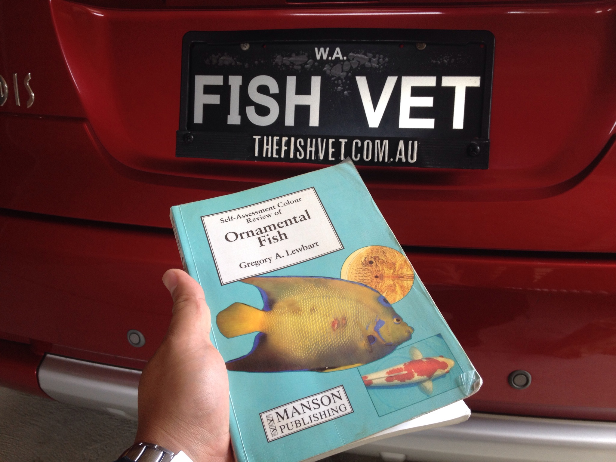
One of my friends gave me this book as a present (one of the best gifts by far, thank you). In this book are 265 cases of fish health problems with diagnostic work ups and solutions to the ailments. This “Self-Assessment Colour Review of Ornamental Fish” by Dr Greg Lewbart is a most excellent publication. It quickly gave me the confidence that I have seen or read about the most common issues that are out there. I highly recommend it if you’re at all serous about being a fish veterinarian.

What’s more is that they’re publishing a second edition (yay!); and they’ve asked me to contribute some of my cases (yay x2!).

Keep a look out for the new edition when it’s ready!

IMG_5247-0.JPG