Buoyancy disorders of ornamental fish: a review of cases seen in veterinary practice.


A great article by Dr William Wildgoose regarding the perennial problem of upside down goldfish.

For the short version, excerpts from the abstract is pasted below:

There were almost equal numbers of short-bodied and long-bodied goldfish. In many cases, there were no external lesions and no consistent environmental factors could be identified… Several radiographic abnormalities were found in goldfish including over-inflation (44%), displacement (22%), fluid accumulation (12%) and rupture (2%) of the swim bladder and intestinal tympany (6%)… There was a very poor response to treatment and several environmental, medical and surgical approaches are discussed.

For the long version, click on this link.


Fish-e Trivia for Monday-itis. Where do Bettas come from?

Did you know that the Siamese fighting fish comes from Siam? Siam is the old name for Thailand. As such, they are tropical fish and the water in which they are kept should reflect that (i.e. 24-28 degrees celsius). Today, the fish is bred in captivity virtually anywhere in the world, provided you can maintain such tropical water conditions for them. The majority of those you purchase from the local fish shop would be from south-east Asia.

On another note, if your Betta is looking a bit lethargic or off its feed, it may be because the water is much too cold for it. Obtain advice from your fish veterinarian or local fish shop about giving them a good healthy environment.

20120818-092345.jpg

Survival after being gutted! The sea turd’s story.

Aquaculture
Volume 359, Number 5 (August 2012)
Growth, metabolism and immune responses to evisceration and the regeneration of viscera in sea cucumber, Apostichopus japonicus
Authors: Yuanqi Zang, Xiangli Tian, Shuanglin Dong, Yunwei Dong
Author Affiliations:
no affiliations available
Source: Aquaculture, Volume 359, Number 5 (August 2012)
Page Numbers: 50 – 60
Available Full Text:
Full Text: Subscription Required to view full text
Format: PDF
Size: Unknown
Location: Publisher’s Site
Authentication: Publisher’s Site
Abstract: Growth, metabolism and immune responses of sea cucumber, Apostichopus japonicus, following evisceration induced artificially, were investigated in this study. The body weight, specific growth rate (SGR), oxygen consumption rate (OCR), activities of immunoenzymes including acidic phosphatase (ACP), alkaline phosphatase (AKP) from hydrolytic system, and catalase (CAT), superoxide dismutase (SOD), total antioxidant capacity (T-AOC) and malonyl-dialdehyde (MDA) from antioxidant system in muscle, intestine and respiratory tree were measured to evaluate the physiological responses of A. japonicusto evisceration and regeneration of viscera. The results showed that body weight of sea cucumber significantly dropped following evisceration and then increased gradually with digestive function resumed. Accelerated growth rates were observed in the regeneration group from the 10th day to 20th day, but there was no significant difference between the regeneration group and control group at the end of the experiment. OCR reduced rapidly after evisceration and increased gradually afterwards. Compared to the control, no significant difference in OCR was found on the 45th day (P0.05). The immune responses of A. japonicuswere highly tissue-specific during the regeneration of viscera. Both ACP and AKP activities in muscle peaked on the 10th day after evisceration, then decreased gradually to normal on the 45th day (P0.05), while those in the regenerated tissues exhibited a differential rising trend from the 20th day after evisceration. SOD activity in muscle was not significantly influenced by evisceration; however, in the regenerated tissues it was enhanced and significantly higher than those in the control on the 45th day (P<0.01). CAT activity in muscle of the regeneration group was significantly higher than in the control during the experiment (P<0.05). Comparatively, CAT activity in the respiration tree increased invariably after evisceration, while in the intestine CAT activity was not significantly affected by evisceration (P0.05). Significant increase was found in both T-AOC and MDA in all three tissues after evisceration (P<0.05); however, they all returned to normal at the end of the experiment. Results of this study indicated that growth, metabolism and immune responses including hydrolytic and antioxidant enzymes in sea cucumber were significantly influenced by evisceration. Although the regeneration group did not catch up with the control in body weight, SGR, OCR, major non-specific immune parameters resumed to normal level within 45days after evisceration, indicating that A. japonicusmight have recovered physiologically from evisceration. Results from this study first presented comprehensive and valuable data of physiological responses to evisceration and the regeneration of viscera in A. japonicus, which would have important implications for those farmers engaged in sea cucumber culture.
Citation: Yuanqi Zang, Xiangli Tian, Shuanglin Dong, Yunwei Dong . Growth, metabolism and immune responses to evisceration and the regeneration of viscera in sea cucumber, Apostichopus japonicus. Aquaculture, Volume 359, Number 5 (August 2012), pp. 50-60, <http://ejournals.ebsco.com/direct.asp?ArticleID=469494601EB864391BCF&gt;
URL: http://ejournals.ebsco.com/direct.asp?ArticleID=469494601EB864391BCF

Daughterless carp project.

We really should be supporting the channelling of funds towards the  “daughterless” carp project instead, see link –
http://www.abc.net.au/landline/content/2012/s3550676.htm

Natural plant extracts to combat Saprolegnia fungus.

Sounds promising!


Aquaculture
Volume 359, Number 5 (August 2012)
The efficacy of selected plant extracts and bioflavonoids in controlling infections of Saprolegnia australis(Saprolegniales; Oomycetes)
Authors: S. Caruana, G.H. Yoon, M.A. Freeman, J.A. Mackie, A.P. Shinn
Author Affiliations:
no affiliations available
Source: Aquaculture, Volume 359, Number 5 (August 2012)
Page Numbers: 146 – 154
Available Full Text:
Full Text: Subscription Required to view full text
Format: PDF
Size: Unknown
Location: Publisher’s Site
Authentication: Publisher’s Site
Abstract: Saprolegniainfections can account for the significant loss of salmonid eggs and adults in breeding condition and although there are a number of chemical agents available to control infections in hatcheries, treatment is difficult and there are reservations regarding their efficacy, prompting a search for suitable replacements. Here, the efficacy of 24 crude plant extracts and 3 bioflavonoids on the growth of Saprolegnia australis were assessed using an in vitro agar diffusion method against a set of synthetic chemotherapeutants commonly used within the aquaculture industry. The 12 best extracts inhibiting growth at =100ppm, were then added to cultures of S. australisin a GP broth and the daily growth was monitored spectrophotometrically over a 7-day period. All 12 plant extracts reduced growth, however, only Atractylodes macrocephala, Zingiber officinale seed, Chrysanthemum and Yucca were as effective as 10ppm malachite green in slowing growth. A subsequent trial, where doses of the plant extract were given at 72 and 120h post-inoculation, found that Rumex obtusifolius, Sophora flavescens, Echinaceaand Z. officinale were the most effective compounds impacting growth. The study demonstrates that certain plant extracts impact on the growth dynamics of S. australisbut further research is required to assess their performance in vivo.
Citation: S. Caruana, G.H. Yoon, M.A. Freeman, J.A. Mackie, A.P. Shinn . The efficacy of selected plant extracts and bioflavonoids in controlling infections of Saprolegnia australis(Saprolegniales; Oomycetes). Aquaculture, Volume 359, Number 5 (August 2012), pp. 146-154, <http://ejournals.ebsco.com/direct.asp?ArticleID=4010BD15760B69FA953A&gt;
URL: http://ejournals.ebsco.com/direct.asp?ArticleID=4010BD15760B69FA953A

Dr Loh, The Fish Vet, in print media.

Check out the interesting articles on my Facebook "Fin Page" – http://www.facebook.com/media/set/?set=a.403427183039570.88397.188036301245327&type=3&l=12842bc857


Follow me on: Facebook "Fin Page" LinkedinYouTubeTwitterBlog
Yours sincerely,

Dr Richmond Loh
BSc, BVMS, MPhil (Vet Path), MANZCVS (Aquatics), MANZCVS (Pathobiology), DipPM.
Veterinarian | Adjunct Senior Lecturer Murdoch University | WAVMA Communications Committee Member |
Secretary Aquatic Animal Health Chapter – Australian and New Zealand College of Veterinary Scientists (ANZCVS)

The Fish Vet, Perth, Western Australia. Mobile Veterinary Service for fish and other aquatic creatures.
http://www.thefishvet.com.au
Ph: +61 (0)421 822 383

Did you know that koi herpesvirus can infect and cause disease in other species?

I know that most herpesviruses are relatively host-specific, however there are a few exceptions – the simian cercopithecine herpes B virus (which can infect monkeys and humans) and Aujeszky’s disease virus (which affects pigs and a wide range of other animals including cattle, sheep, goats, dogs, cats, mink, foxes, raccoons and rats). Malignant catarrhal fever is another herpesvirus that when it jumps from its natural sheep host to infect cattle, produces severe acute disease that is almost always fatal!

Recent research has demonstrated that although KHV doesn’t cause overt disease in other hosts, it IS capable of infecting them. After mentioning all those other diseases previously, it makes me wonder if under certain circumstances, the KHV may produce disease in other species it is capable of infecting (e.g. viral mutation, immunosuppression, environmental thermal changes, etc). Think about gallid herpesvirus 2 (Marek’s disease virus) picked up an oncogene somewhere along its evolution (M. Bennett, pers. comm).

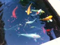
Dr Sven Bergman and co. in Germany have been working on KHV and recently, they found that not only were carp dying, but it could also infect tench and roach! This information will soon be published and we are privileged to be among the first to know.

The information below are extracts from scientific papers published by Dr Sven Bergman and co. in Germany (provided with permission from the author).

I have forwarded these papers to the lead Australian scientist, Dr Ken McCol, who is planning to inflict this virus on carp and the like, to appeal to his better judgement to abandon his project.

The key sentences to support the prevention of KHV release in Australia are in bold.

How Host Specific is Infection with Koi Herpesvirus (KHV) for Real?
S.M. Bergmann1*, J. Kempter2, D. Fichtner1
1 Friedrich-Loeffler-Institut, Federal Research Institute for Animal Health, Institute of Infectology,
Sudufer 10, 17493 Greifswald-Insel Riems, Germany (* sven.bergmann@fli.bund.de)
2 Agricultural University, Department of Aquaculture, K. Krolewicza 4, 71-550 Szczecin, Poland

Key words: KHV infection, detection methods, nested PCR, non Cyprinus carpio species
Contrary to the theory of host specificity of herpesviral infections, channel catfish herpesvirus
(CCV) is able to infect more than one species, e.g., blue catfish (Ictaluris furcatus), channel catfish
(Ictaluris punctatus), and white catfish (Ictaluris catus). Although African catfish (Clarias
gariepinus), Asian catfish (Clarias batrachus), and other species are resistant to clinical CCV infection,
CCV infected a mammalian cell line obtained from Hawaiian monk seal (Monachus schauinslandi).
Due to these findings, we sought more host species in the framework of koi herpesvirus
(KHV) infection (KHVI). KHV disease (KHVD) occurs only in Cyprinus carpio (common carp and
koi). Among other species, we tested goldfish (Carassius auratus), crucian carp (C. carassius),
tech (Tinca tinca), grass carp (Ctenopharyngodon idella), bighead (Aristichthys nobilis), silver carp
(Hypophthalmichthys molitrix), sheatfish (Silurus glanis), sturgeon species (Acipenser gueldenstaedtii,
A. oxyrhynchus, A. ruthenus), and ornamental fish by different PCRs. All non Cyprinus
carpio fish had never expressed any clinical sign of KHVD. Using different methods for DNA
extraction (clumbs, fluid reagents) from different organs (organs, swabs, smears, blood, etc.), KHV
DNA was detected in most of the experimentally or naturally-infected fish by nested PCR. Positive
PCR results were confirmed by in situ hybridization using different probes, by sequencing of PCR
products, or by immunofluorescence assay using polyclonal and monoclonal antibodies developed
against KHV. In the same framework we tried to establish and test the sensitivity of non-lethal sampling
methods, e.g., separation of leukocytes or gill swabs. Our aim was to exclude or detect KHVI
in fish (carriers) that were in contact with KHV-infected carp or other fish affected by KHVI.

The Israeli Journal of Aquaculture – Bamidgeh 61(3), 2009
Detection of koi herpes virus (KHV) genome in apparently healthy fish.
S. M. Bergmann1*, H. Schütze1, U. Fischer1, D. Fichtner1,

M. Riechardt1, K. Meyer3, D. Schrudde1 and J. Kempter2

1 Friedrich-Loeffler-Institut, Federal Research Institute for Animal Health, Institute of Infectology,
Südufer 10, 17493 Greifswald-Insel Riems, Germany; 2 Agricultural University, Department of
Aquaculture, K. Królewicza4, 71-550 Szczecin, Poland; 3 Veterinary High school, Hannover, Bünteweg
17, 30559 Hannover, Germany

Abstract
Koi herpesvirus (KHV) induces a lethal disease in species belonging to Cyprinus carpio, covering
common carp and koi or fancy carp. To date, other cyprinid fish species kept together with KHV
infected carp or koi, such as goldfish (Carassius auratus) or grass carp (Ctenopharyngodon idella) had
never shown any sign of KHV infection. Unexplainable outbreaks of KHV infection in common
carp or koi led to the suspicion that more disease influencing factors exist, than so far explained. In
challenge experiments and by natural routes of infection, it has been demonstrated that naive carp
or koi can be infected by the following mechanisms; exposure to water from severe KHV diseased
fish, co-habitation with KHV infected carp, by injection or immersion with cell supernatant of KHV
infected cultures. Experiments were conducted to determine if apparently healthy koi (carriers/
survivors) or non-Cyprinus carpio species stocked with KHV infected carp could become infected.
The study proved that these species were able to transfer KHV to naïve carp. As a result scientists
suspected that some ornamental fish species could act as reservoirs of KHV infection.
In this study goldfish, grass carp, blue back ide (Leuciscus idus) and Ancistrus sp. were screened
with routinely used diagnostic methods such as virus isolation in cell cultures and PCR and found
to be KHV negative. This suggested that these fish species could not be infected by KHV. However,
when using a more sensitive nested PCR, KHV DNA was detected from some of these fish. As
confirmative methods sequence analysis of the nested PCR products and in-situ hybridization with
different KHV probes were used. This study showed that the following cyprinid species; goldfish,
grass carp and ide, and also non-cyprinid species Ancistrus sp. may act as a carrier of KHV.

Bull. Eur. Ass. Fish Pathol., 29(5) 2009, 145

KOI HERPES VIRUS: DO ACIPENSERID RESTITUTION PROGRAMS POSE A THREAT TO CARP FARMS IN THE DISEASE-FREE ZONES?
Jolanta KEMPTER 1*, Jacek SADOWSKI 1, Heike SCHÜTZE 2, Uwe FISCHER 2, Malte
DAUBER 2, Dieter FICHTNER 2, Remigiusz PANICZ 1, and Sven M. BERGMANN 2

1West Pomeranian University of Technology, Division of Aquaculture, Szczecin, Poland
2 Friedrich-Loeffler-Institut, Federal Research Institute for Animal Health, Suedufer 10,
17493 Greifswald-Insel Riems, Germany
Kempter J., Sadowski J., Schütze H., Fischer U., Dauber M., Fichtner D., Panicz R., Bergmann S.M. 2009.

Background. Sturgeons have long been extinct in Polish inland waters. A substantial effort has recently been put
into their restitution, covering the drainage areas of two major Polish rivers, the Oder and the Vistula. The
stocked fishes are clinically healthy, but very little is known about their potential to transmit viral diseases including
koi herpes virus (KHV) to healthy fishes of other species, which may pose a threat to the disease-free zones.
This study was intended to determine if sturgeons could be asymptomatic carriers of KHV.
Materials and Methods. A total of 29 sturgeons (two species; length 8–37 cm) originating from fish farms in
northern Poland with a known KHV history in common carp or koi in the area were examined: 15 Russian sturgeons,
Acipenser gueldenstaedtii, with clinical signs of a disease and 14 asymptomatic Atlantic sturgeons,
A. oxyrinchus. The former were sent to the laboratory alive while the latter were sent fixed in ethanol. As it is
required for detection of a latent KHV infection in acipenserids, two independent procedures were applied. The
preliminary results were obtained using PCR. Those findings were subsequently confirmation by nested PCR. The
latter procedure consists of sequence analysis of PCR products and direct detection of KHV infected cells in tissue
materials by in-situ hybridization on nucleic acid level or indirect immunofluorescence on KHV protein level.
Results. KHV genome parts were found in nine Russian sturgeons and four Atlantic sturgeons. Comparison of
PCR results obtained from three primer pairs used for KHV diagnostic in sturgeon showed that those designed
by Bercovier et al. (2005) were most sensitive and robust for this purpose. In order to confirm the presence of
viral particles the most useful method was in-situ hybridization (ISH), allowing the detection of KHV in gill samples
obtained from live sturgeons.
Conclusion. This preliminary study shows that sturgeons can be carriers of KHV. Therefore a viral diagnostics
is highly recommended not only for sturgeons obtained from the environment but also for fertilized eggs, fry, and
fish intended for re-stocking measurements of inland waters.
Keywords: Koi herpesvirus, Acipenser gueldenstaedtii, Russian sturgeon, Acipenser oxyrinchus, Atlantic sturgeon,
PCR, nested PCR, ISH, iIFA

ACTA ICHTHYOLOGICA ET PISCATORIA (2009) 39 (2): 119–126

20120810-075121.jpg

A pat on the back for the World Aquatic Veterinary Medical Association.

This was an impromptu email to the World Aquatic Veterinary Medical Association (WAVMA) from a new veterinary graduate, commending the WAVMA (and my book!) on the great work that’s being done in progressing aquatic veterinary medicine.


From: Chad Harris
Date: Sat, Aug 4, 2012 at 9:49 PM
Subject: Thank you all!
To: WAVMA

WAVMA Members,
I am a recent graduate from Ross University practicing in Austin, Texas. I have been following your posts and reading TONS of material, including Dr. Loh’s book, which has given me a wealth of “just scratching the surface” knowledge. As we all know, it is unfortunate that we do not get more exposure to fish/aquatic medicine in school. Long story short, I am attempting to bring fish medicine and surgery into my practice and I saw my first patient, Leo the fancy lionhead goldfish yesterday. Thanks to all of your posts, I didn’t skip a beat and Leo is doing much better today! If any of you have any tips/tools of the trade to send my way, by all means don’t hesitate.

Thank you so much,

Dr. Chad Harris, DVM
North Austin Animal Hospital
Austin, TX


Check out WAVMA by clicking on this link.

Check out Dr Loh’s books by clicking on this link.


20120810-074541.jpg

What does it mean if fish have “dropsy”?

Dropsy is not a disease diagnosis, but is actually a descriptive term for the clinical signs observed in fish. The term has often been misused as being diagnostic for bacterial disease, when all it actually means is an abnormal accumulation of fluid in a body cavity or in the subcutaneous tissues. Another medical term that can be used is anasarca. Grossly, the scales of the fish will protrude from the body (due to subcutaneous oedema), giving them the classic “pine cone” appearance. Fluid may also accumulate in the coelomic cavity, giving them a “pot-belly” appearance. This condition is limited to fishes in a freshwater environment and the reason for this is explained in the following sentence. Fishes that live in freshwater are in a hypo-osmotic environment. And so, if there are any severe external lesions (e.g. large skin ulcers from bacterial infection or severe gill damage from fungal infections), there could be a net influx of fluid into the body. If the kidneys are damaged such that normal excretion of urine is reduced, dropsy can also result. The prognosis is poor and such fish should be euthanased. It is useful to examine the diseased fish immediately post-euthanasia to identify the reason for the disease to prevent it from occurring in its tankmates.

To summarise, the cause of dropsy is not limited to bacterial infection, but it can occur as a result of skin disease, gill disease, renal disease or congestive heart failure which can have a variety of aetiologies.


20120810-074429.jpg