What other protein sources are available for making fish food?

Although ‘mad cow disease’ should not be an issue in fish, it is still good to take precaution and not use it in primary food production. But what about proteins from other species? What is their fate? Usually these go to waste, but should it really be that way?

Learn more at this link:
Non-ruminant protein as fish feed.

How do you know if there’s sufficient oxygen in your pond?

Due to the lack of photosynthetic activity by plants while there is no sunlight, my suggestion is for you to wake up at the break of dawn and check your fish pond. In a healthy balanced system, the fish should not be gasping for air.

Below is a picture of a healthy pond, at the break of dawn.

20130221-063145.jpg

New recreational fishing guidelines for Western Australia.

Bag and size limits help ensure our aquatic resources remain sustainable for future generations. Bag limits assist in sharing the resource and contribute to the sustainable
management of the State’s fish stocks. Size limits allow fish to reach maturity to complete their breeding cycle.

The new rules came into effect on 1 February 2013.

Learn more by following this link – http://www.fish.wa.gov.au/Fishing-and-Aquaculture/Recreational-Fishing/Recreational-Fishing-Rules/Pages/default.aspx

What do you think it means if crabs avoid getting electric shock?

Have a read of this article and let me know your thoughts – http://blogs.smithsonianmag.com/science/2013/01/new-study-suggests-crabs-can-feel-pain-after-all

What did Dr Loh learn at AquaVet II?

I believe you may have read about bits and pieces when I was attending AquaVet. This is the full report of my learnings and my recommendations.

There are bits in there relevant to veterinarians, educators, government, industry and ornamental fish owners.

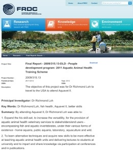
You can access my report for the FRDC- funded trip at this link:
http://frdc.com.au/research/final-reports/Pages/2009-315-13-DLD.aspx

20130218-115201.jpg

FYI, the intensive timetable for the learnings are provided below:

Aquavet II Schedule for 2012

Sunday, 27. May 2012

14:00 Orientation – Directors

15:00 RWU Required Safety Lecture – Caitlin Conley

16:00 Fish Haematology- Dr. Diane Brown – Harvard Medical School

18:30 Fish Histology (continued) – Dr. Diane Brown

Monday, 28. May 2012

8:00 Corals – Dr. Ilze Berzins – IAAAM President

13:00 Corals – continued – Dr. Ilze Berzins

15:00 Comparative Anatomy of Shellfish – Introduction to Pathology of Molluscan Diseases – Dr. Roxanna

18:30 Invertebrate Pathology – Diseases of Bivalves – Dr. Roxanna Smolowitz

Tuesday, 29. May 2012

8:00 Using Animals in Research – Dr. Amy Hancock-Ronemus

9:30 Introduction to Diseases of Aquaculture Species – Warmwater – Catfish – Dr. Sherman Jack

13:00 Pathology of Catfish Diseases – Dr. Sherman Jack

18:30 Diagnostic Case Studies and Practicum – Aquacultured Species – Drs. Sherman Jack and Rod Getchell

Wednesday, 30. May 2012

8:00 Toxicologic Pathology of Fishes – Dr. Jeff Wolf.

13:00 Toxicologic Pathology of Fishes (continued) – Dr. Jeff Wolf

13:00 Toxicologic Pathology of Fishes (continued) – Drs. Jeff Wolf and Rod Getchell

Thursday, 31. May 2012

8:00 Toxicologic Pathology of Fishes (continued) – Dr. Jeff Wolf

13:00 Toxicologic Pathology of Fishes (continued) – Dr. Jeff Wolf

18:30 Toxicologic Pathology of Fishes (continued) – Drs. Jeff Wolf and Rod Getchell

Friday, 1. June 2012

8:00 Invertebrate Pathology – Diseases of Bivalves – Dr. Roxanna Smolowitz

13:00 Normal Anatomy and Diseases of Cephalopods and Opistobranchs – Dr. Roxanna Smolowitz

18:00 Normal Anatomy of Echinoderms and Limulus – Dr. Roxanna Smolowitz

Saturday, 2. June 2012

8:00 Parasites in Aquatic Animals – Dr. Sarah Poynton

Sunday, 3. June 2012 – OFF

8:30 leave campus for Whale Watch – Barnstable, MA

Monday, 4. June 2012

8:00 Invertebrate Pathology – WET LAB – Dr. Roxanna Smolowitz

13:00 Invertebrate Pathology – WET LAB – Dr. Roxanna Smolowitz

18:30 open

Tuesday, 5. June 2012

8:00 Diagnostic Case Studies and Practicum – Aquacultured Species – Dr. Mark Fast, Dr. Sal Frasca and Dr. Rod Getchell

11:00 Diseases of Coldwater Aquaculture Species – Infectious and Non-Infectious – Drs. Mark Fast, Sal Frasca, and Rod Getchell

13:00 Diseases of Coldwater Aquaculture Species – Infectious and Non-Infectious (continued) – Drs. Mark Fast, Sal Frasca, and Rod Getchell

18:30 Diseases of Coldwater Aquaculture Species – Infectious and Non-Infectious (continued) – Drs. Mark Fast and Rod Getchell

Wednesday, 6. June 2012

8:00 Conundrums – Drs. Mark Fast, Rod Getchell and/or Paul Bowser

10:00 Fish as Lab Animals – Dr. Paul Bowser

13:00 Normal Anatomy of Crustaceans – Dr. Roxanna Smolowitz

18:30 Diseases of Crustaceans – Dr. Roxanna Smolowitz

Thursday, 7. June 2012

8:00 Neoplasia of Fish – Dr. Renate Reimschuessel

13:00 Fish Diagnostics and Techniques WET LAB – Dr. Rod Getchell

18:30 Emerging Viral Fish Diseases in the US – SVCV, SHSV, KHV, LMBV – Dr. Rod Getchell

Friday, 8. June 2012

8:00 Overview of the Principal Infectious Diseases Found in Farmed Penaeid Shrimp – Dr. Arun Dhar and Dr. Robert Bullis

13:00 Overview of the Principal Infectious Diseases Found in Farmed Penaeid Shrimp – Drs. Arun Dhar and Robert Bullis

Saturday, 9. June 2012

By 12:00 check out

Green water for larval fish rearing, but what’s in it?

This article is almost a leaf out of the koi club book on fish breeding and larval rearing! What do koi breeders do at the moment? Preparation of the pond can take 2 weeks to a month. Some introduce medium sized koi to the fry pond to consume any of the large predatory insect larvae. Approximately one week prior to introducing the fertilised eggs to the pond, the large fish are removed and the pond is fertilised. This will help establish ‘green water’. What’s in this ‘green water’? Is it just algae? Are there bugs in there that fish eat?
Read on to find out more.

Aquaculture Research
Volume 44, Number 3 (February 2013)
Experimental evaluation of inorganic fertilization in larval giant grouper (Epinephelus lanceolatus) production

Authors: Kwee Siong Tew, Pei-Jie Meng, Hsi-Sheng Lin, Jian-Hua Chen, Ming-Yih Leu
Author Affiliations:
no affiliations available
Source: Aquaculture Research, Volume 44, Number 3 (February 2013)
Page Numbers: 439 – 450
Available Full Text:
Full Text: Subscription Required to view full text
Format: PDF
Size: unknown
Location: Publisher’s Site
Authentication: EBSCOhost EJS

Abstract: A major constraint in successful larviculture of groupers has been the small gape of the larvae and hence the requirement for small prey at first feeding. In this study, we examined how maintaining a phosphate concentration of 100 µg P L-1and an inorganic nitrogen (N) level of 700 µg N L-1 via weekly fertilization with inorganic fertilizers affected phytoplankton, zooplankton and giant grouper larval survival in relation to a control group that was provided with rotifers immediately after larvae hatched. Unicellular algae, zooplankton within the size ranges of 10–50 µm and 50–100 µm and survival of giant grouper larvae were all significantly higher in the fertilized treatment compared with the control. Stomach analysis revealed that ciliates and flagellates were actively consumed by larval fish in the fertilized group, whereas few rotifers were consumed in the control. We conclude that the inorganic fertilization method provides high densities of suitable-sized prey for larval groupers at the onset of exogenous feeding before they are able to consume larger, commercially available rotifers and copepods.
Citation: Kwee Siong Tew, Pei-Jie Meng, Hsi-Sheng Lin, Jian-Hua Chen, Ming-Yih Leu . Experimental evaluation of inorganic fertilization in larval giant grouper (Epinephelus lanceolatusBloch) production. Aquaculture Research, Volume 44, Number 3 (February 2013), pp. 439-450,
URL: http://ejournals.ebsco.com/direct.asp?ArticleID=454495BEFA1675A435E7


Yours sincerely,

Dr Richmond Loh
BSc, BVMS, MPhil (Vet Path), MANZCVS (Aquatics), MANZCVS (Pathobiology), DipPM.
Veterinarian | Adjunct Senior Lecturer Murdoch University | WAVMA President-Elect |
Secretary Aquatic Animal Health Chapter – Australian and New Zealand College of Veterinary Scientists (ANZCVS)
The Fish Vet, Perth, Western Australia, AUSTRALIA.
Mobile Veterinary Medical & Diagnostic Services for fish and other aquatic creatures.
http://www.thefishvet.com.au
Ph: +61 (0)421 822 383

Are you getting what you paid for in fish food?

There are has been a great deal of research into substituting fish proteins with plant proteins, but I don’t believe we’ve gotten to the bottom of it yet. Also, did you know that chicken used to be a good source of omega 3’s when they were truly free ranging and their diet consisted if insects and other invertebrates? Fish are currently held as being good sources of omega 3’s, but what will happen to this if their diet is changed?

And so, it is not a good idea for manufacturers of fish food to be substituting. It does the industry harm and an unprofitable fish farm will eventually lead to unprofitable feed manufacturers as a flow on effect.

Learn more by clicking on the link below:

Fake fish feed.

Salmon more slimy on top!

I had always noticed a difference in the frequency of mucus cells over the surface of the fish through my work, and now we have a report that quantifies it!

Journal of Fish Diseases
Volume 36, Number 2 (February 2013)
Body site matters: an evaluation and application of a novel histological methodology on the quantification of mucous cells in the skin of Atlantic salmon, Salmo salar L.
Authors: K Pittman, A Pittman, S Karlson, T Cieplinska, P Sourd, K Redmond, B Ravnøy, E Sweetman
Author Affiliations:
no affiliations available
Source: Journal of Fish Diseases, Volume 36, Number 2 (February 2013)
Page Numbers: 115 – 127
Available Full Text:
Full Text: Subscription Required to view full text
Format: PDF
Size: unknown
Location: Publisher’s Site
Authentication: EBSCOhost EJS
Abstract: Mucous cell size and distribution were investigated in the skin of five salmon using a novel stereology-based methodology: one (48 cm) fish to test 15 tissue treatment combinations on measures of cell area and density on the dorsolateral region and, using the most suitable treatment, we mapped mucous cell differences between body regions on four (52 cm) salmon, comprising a male and a female on each of two diets. The section site, decalcification, embedding medium and plane of sectioning all impacted significantly on mucous cell size, whereas mucous cell density is more robust. There were highly significant differences in both mucosal density and mean mucous cell size depending on body site: the dorsolateral skin of the four salmon had significantly denser (about 8% of skin area) and larger (mean about 160 µm2) mucous cells, whereas the lowest mean density (about 4%) and smallest mean area (115 µm2) were found on the head. We found that 100 random measurements may be sufficient to distinguish differences >7 µm2in mean mucous cell areas. The results further suggest that salmon exhibit a dynamic repeatable pattern of mucous cell development influenced by sex, diet and possibly strain and season.
Citation: K Pittman, A Pittman, S Karlson, T Cieplinska, P Sourd, K Redmond, B Ravnøy, E Sweetman . Body site matters: an evaluation and application of a novel histological methodology on the quantification of mucous cells in the skin of Atlantic salmon, Salmo salar L.. Journal of Fish Diseases, Volume 36, Number 2 (February 2013), pp. 115-127, <http://ejournals.ebsco.com/direct.asp?ArticleID=41639E48C3F2C086D053&gt;
URL: http://ejournals.ebsco.com/direct.asp?ArticleID=41639E48C3F2C086D053